Ultrasound Machine: How It Works, Types, Uses, Benefits, Risks, and the Future of Ultrasound Technology

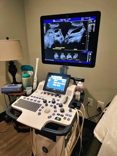
An ultrasound machine is a medical imaging system that uses high-frequency sound waves to create real-time images of structures inside the body. Unlike X-ray and CT imaging, ultrasound does not use ionizing radiation. This makes it an important diagnostic and monitoring technology in medicine, particularly for pregnancy care, abdominal imaging, cardiovascular examinations, emergency medicine, and procedures guided by imaging.
Modern ultrasound systems range from large cart-based machines found in hospitals to compact portable and handheld devices that can connect to tablets or smartphones. Their ability to produce images in real time has made ultrasound valuable not only for diagnosis but also for guiding medical procedures and monitoring changes in the body.
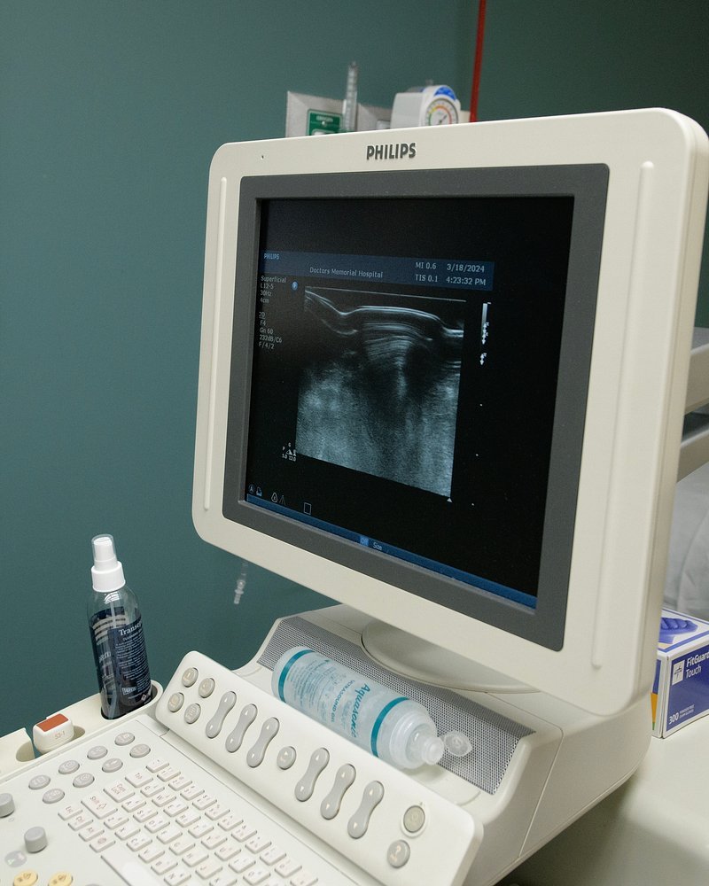
Ultrasound technology is based on a relatively simple physical principle. A transducer sends sound waves into the body. When those waves encounter boundaries between tissues with different acoustic properties, some of the energy is reflected back toward the transducer. The machine measures these returning echoes and processes them into an image.
Table of Contents
The technology has evolved considerably from early static ultrasound systems. Modern equipment can produce two-dimensional images, three-dimensional and four-dimensional images, Doppler measurements of blood flow, elastography measurements of tissue stiffness, and increasingly sophisticated computer-assisted imaging.
What Is an Ultrasound Machine?
An ultrasound machine is a device that uses high-frequency sound waves to visualize internal structures and, in some applications, measure movement or blood flow.
The procedure is also called sonography or ultrasonography.
The system normally consists of several major components:
- Transducer or probe
- Computer and signal-processing system
- Display monitor
- Control panel
- Electrical power system
- Image-storage system
- Software for image processing and measurements
The transducer is one of the most important components because it both sends sound waves into the body and receives echoes returning from tissues.
The machine then processes these signals to construct an image.
What Does Ultrasound Mean?
Ultrasound refers to sound waves with frequencies above the range normally audible to humans.
Humans generally hear frequencies from approximately 20 Hz to 20 kHz, while medical diagnostic ultrasound operates at much higher frequencies, commonly in the megahertz range.
The higher frequency allows ultrasound to provide detailed images of relatively shallow structures, although higher-frequency waves generally do not penetrate as deeply as lower-frequency waves.
This creates an important trade-off between resolution and penetration.
How Does an Ultrasound Machine Work?
The operation of an ultrasound machine can be explained through several stages.
Step 1: The Transducer Produces Sound Waves
The ultrasound transducer contains piezoelectric elements.
When an electrical signal is applied to these elements, they change shape and generate mechanical vibrations.
These vibrations produce ultrasound waves.
Step 2: Sound Waves Enter the Body
The transducer is placed against the skin, usually with ultrasound gel between the probe and skin.
The gel eliminates air between the transducer and skin because air interferes strongly with the transmission of ultrasound.
The sound waves travel into the body.
Step 3: Waves Encounter Tissues
As ultrasound travels through the body, it encounters different tissues.
Some of the sound energy continues deeper into the body, while some is reflected back toward the transducer.
The amount of reflection depends partly on differences in acoustic impedance between tissues.
Step 4: The Transducer Receives Echoes
The same transducer or its elements detect returning echoes.
The piezoelectric elements convert mechanical vibrations back into electrical signals.
Step 5: The Computer Processes the Signals
The ultrasound system analyzes the returning signals.
It determines information such as:
- How long an echo took to return
- How strong the echo was
- Where the echo originated
- How tissues are moving
- How blood is flowing
Step 6: An Image Is Created
The computer converts the processed information into an image displayed on the monitor.
Because the process occurs rapidly, ultrasound can produce images in real time.
What Is an Ultrasound Transducer?
The transducer, commonly called a probe, is the component that comes into direct contact with the patient’s body.
It performs two critical functions:
- It sends ultrasound waves into the body.
- It receives returning echoes.
Different probes are designed for different applications.
A probe used for a pregnancy examination is shaped differently from one designed for cardiac imaging or superficial structures.
Types of Ultrasound Transducers
Linear Transducer
A linear probe has a flat footprint and is commonly used for structures close to the skin.
Applications include:
- Blood vessels
- Thyroid
- Breast
- Tendons
- Muscles
- Skin
- Peripheral nerves
Linear probes generally operate at relatively high frequencies, providing good resolution for superficial structures.
Curvilinear Transducer
A curvilinear or curved-array probe has a curved surface.
It provides a wider field of view and is frequently used for deeper structures.
Applications include:
- Abdominal imaging
- Obstetric imaging
- Pelvic examinations
Phased-Array Transducer
A phased-array probe is commonly used for cardiac imaging.
Its small footprint allows it to work through narrow spaces between ribs.
The ultrasound beam can be electronically steered, allowing the system to examine the heart from different angles.
Endocavitary Transducer
Endocavitary probes are designed for examinations in which the transducer is inserted into a body cavity.
Examples include:
- Transvaginal ultrasound
- Transrectal ultrasound
They allow detailed imaging of structures that may be difficult to visualize effectively from outside the body.
Specialized Probes
Other probes can be designed for applications such as:
- Intravascular ultrasound
- Endoscopic ultrasound
- Intraoperative ultrasound
- Musculoskeletal imaging
- Pediatric imaging
Ultrasound Frequencies
Ultrasound probes operate across different frequency ranges.
A higher-frequency probe generally provides better spatial resolution but does not penetrate as deeply.
A lower-frequency probe generally penetrates deeper but produces lower resolution.
This relationship means sonographers choose the transducer according to the body structure being examined.
For example, a superficial thyroid examination may use a high-frequency linear probe, while an abdominal examination may require a lower-frequency curved probe.
Types of Ultrasound Imaging
Ultrasound is not a single imaging mode.
Modern machines can produce several types of information.
2D Ultrasound
Two-dimensional ultrasound is the conventional form of sonography.
It creates flat images showing the shape and internal structure of tissues.
It remains one of the most widely used ultrasound techniques.
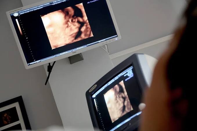
3D Ultrasound
Three-dimensional ultrasound combines multiple image slices to create a volumetric representation.
It can be particularly useful in obstetrics and certain examinations where three-dimensional anatomy provides additional information.
4D Ultrasound
Four-dimensional ultrasound is essentially three-dimensional imaging displayed over time.
The fourth dimension is time.
This allows clinicians to observe movement of three-dimensional structures.
Doppler Ultrasound
Doppler ultrasound evaluates movement, particularly the movement of blood.
The Doppler effect describes changes in the observed frequency of sound waves when the source or reflecting material is moving.
In medical ultrasound, the technique can provide information about blood-flow direction and velocity.
Color Doppler
Color Doppler displays blood-flow information using colors overlaid on the grayscale image.
It can help clinicians visualize blood movement through vessels and the heart.
Spectral Doppler
Spectral Doppler presents blood-flow velocity information as a waveform.
It can help clinicians measure characteristics such as peak velocities and flow patterns.
Power Doppler
Power Doppler is sensitive to blood-flow signals and can be useful for detecting relatively low-flow states, although it does not provide the same directional information as conventional color Doppler.
Ultrasound in Pregnancy
Pregnancy is one of the best-known applications of ultrasound.
Obstetric ultrasound can be used to evaluate:
- Fetal development
- Fetal anatomy
- Fetal position
- Placental location
- Amniotic fluid
- Fetal heart activity
- Growth measurements
- Multiple pregnancies
Ultrasound can also help estimate gestational age and monitor fetal growth.
The American College of Obstetricians and Gynecologists describes ultrasound as an important tool in pregnancy care and notes that standard diagnostic ultrasound has an established record of use when appropriately performed. (acog.org)
However, ultrasound should be performed for appropriate medical reasons and by trained professionals rather than being treated simply as a souvenir or entertainment service.
Ultrasound for Abdominal Imaging
Abdominal ultrasound can examine organs such as:
- Liver
- Gallbladder
- Kidneys
- Pancreas
- Spleen
- Abdominal blood vessels
It can help detect or investigate conditions such as gallstones, cysts, fluid collections, organ enlargement and certain masses.
One advantage is that ultrasound can provide real-time imaging without ionizing radiation.
Ultrasound for the Heart
Cardiac ultrasound is called echocardiography.
An echocardiogram uses ultrasound to visualize the heart.
It can provide information about:
- Heart chambers
- Heart valves
- Heart muscle movement
- Blood flow
- Pumping function
- Fluid around the heart
Doppler techniques are particularly important in echocardiography because they allow clinicians to evaluate blood movement through the heart and vessels.
Ultrasound for Blood Vessels
Vascular ultrasound can examine arteries and veins.
Duplex ultrasound combines conventional grayscale imaging with Doppler information.
It can help evaluate blood flow and identify abnormalities such as narrowing or obstruction in certain blood vessels.
It is commonly used in examinations of the:
- Carotid arteries
- Leg veins
- Peripheral arteries
Ultrasound in Emergency Medicine
Emergency departments increasingly use point-of-care ultrasound, or POCUS.
Clinicians can perform focused ultrasound examinations at the bedside to answer specific clinical questions.
For example, ultrasound can help assess:
- Internal bleeding
- Heart activity
- Fluid around the heart
- Lung findings
- Bladder volume
- Abdominal conditions
- Blood vessels
- Certain causes of shortness of breath
Because the machine can be brought directly to the patient, point-of-care ultrasound can be particularly useful in emergency and critical-care environments.
Point-of-Care Ultrasound
Point-of-care ultrasound refers to ultrasound performed and interpreted by clinicians at or near the patient’s bedside as part of immediate clinical assessment.
Portable devices have made this approach increasingly accessible.
Instead of transporting a patient to a radiology department, a clinician can bring a compact ultrasound system directly to the patient.
This can be valuable in:
- Emergency departments
- Intensive care units
- Ambulances
- Operating rooms
- Rural clinics
- Remote medical environments
The FDA has cleared numerous ultrasound systems for medical use, including portable and handheld devices, but appropriate training and clinical interpretation remain important. (fda.gov)
Handheld Ultrasound Machines
Handheld ultrasound devices are among the most significant recent developments in medical imaging.
A handheld device can be small enough to fit into a pocket or medical bag.
Some models connect to smartphones or tablets, while others have integrated displays.
They can provide imaging in locations where conventional ultrasound equipment would be impractical.
Potential uses include:
- Emergency medicine
- Primary care
- Critical care
- Rural healthcare
- Sports medicine
- Obstetrics
- Vascular assessment
- Procedural guidance
The small size does not eliminate the need for appropriate training. A portable machine can produce an image, but correctly interpreting that image still requires clinical knowledge and experience.
Ultrasound for Medical Procedures
Ultrasound is frequently used to guide procedures.
Examples include:
- Needle placement
- Central venous access
- Nerve blocks
- Fluid drainage
- Biopsies
- Injections
- Regional anesthesia
Real-time imaging can help clinicians see the target structure and monitor the position of a needle or instrument.
This can potentially improve precision and reduce complications compared with procedures performed without imaging guidance.
Ultrasound in Musculoskeletal Medicine
Musculoskeletal ultrasound can examine:
- Muscles
- Tendons
- Ligaments
- Joints
- Nerves
- Soft tissues
It is useful because the clinician can observe structures while they move.
For example, a tendon can be examined dynamically while the patient moves the associated joint.
This dynamic capability is difficult to reproduce with some other imaging techniques.
Ultrasound in Breast Imaging
Ultrasound can be used as an additional imaging method in breast assessment.
It can help characterize certain masses and distinguish between cystic and solid structures.
However, ultrasound does not simply replace mammography.
The appropriate imaging approach depends on factors such as age, symptoms, breast characteristics and the clinical question.
The FDA describes breast ultrasound as a supplemental imaging tool in selected situations rather than a universal replacement for mammography. (fda.gov)
Ultrasound in Thyroid Examination
The thyroid is relatively superficial, making it well suited to high-frequency ultrasound.
Thyroid ultrasound can evaluate:
- Thyroid size
- Nodules
- Cysts
- Tissue characteristics
- Nearby lymph nodes
Ultrasound can also be used to guide needle procedures when tissue sampling is required.
Ultrasound in Kidney and Urinary Tract Imaging
Ultrasound can evaluate the kidneys and urinary bladder.
It can help investigate:
- Kidney size
- Cysts
- Urinary obstruction
- Bladder volume
- Certain types of stones
- Fluid accumulation
Because ultrasound does not use ionizing radiation, it can be useful in situations where repeated imaging may be required.
Ultrasound in Liver and Gallbladder Imaging
Ultrasound is frequently used to evaluate the hepatobiliary system.
It can help detect:
- Gallstones
- Gallbladder inflammation
- Bile duct dilation
- Liver abnormalities
- Cysts
- Certain masses
It can also be combined with Doppler techniques to examine blood flow within major vessels.
Ultrasound vs X-Ray
Ultrasound and X-ray use fundamentally different physical principles.
X-rays are electromagnetic radiation.
Ultrasound uses mechanical sound waves.
| Feature | Ultrasound | X-Ray |
|---|---|---|
| Energy type | Sound waves | Electromagnetic radiation |
| Ionizing radiation | No | Yes |
| Real-time imaging | Yes | Usually not |
| Bone imaging | Limited | Excellent |
| Soft tissue imaging | Often useful | More limited |
| Pregnancy use | Common | Used when medically necessary |
| Blood-flow assessment | Doppler capable | Not directly |
| Portability | High | Variable |
The two technologies complement each other rather than serving as universal substitutes.
Ultrasound vs CT Scan
CT uses X-rays to create cross-sectional images of the body.
Ultrasound uses sound waves.
CT generally provides much greater anatomical detail for many deep structures and is less affected by gas and bone.
Ultrasound has the advantages of real-time imaging, portability and lack of ionizing radiation.
The appropriate technology depends on the clinical question.
Ultrasound vs MRI
MRI uses strong magnetic fields and radiofrequency energy to create detailed images.
It generally provides excellent soft-tissue contrast.
Ultrasound is usually faster, more portable and less expensive, and it can provide real-time imaging.
MRI, however, can visualize many structures that are difficult to evaluate adequately with ultrasound.
Again, the two technologies are complementary.
Benefits of Ultrasound Machines
No Ionizing Radiation
Diagnostic ultrasound does not use ionizing radiation.
This is one of its most important advantages.
The FDA explains that ultrasound imaging does not use ionizing radiation and has a long history of safe medical use when appropriately performed. (fda.gov)
Real-Time Imaging
Ultrasound can show movement in real time.
This is particularly useful for the heart, blood flow and procedures.
Portable
Modern ultrasound systems can be moved between departments, while handheld devices can be taken directly to patients.
Relatively Accessible
Ultrasound equipment can be less expensive than large CT or MRI systems, although advanced systems can still be costly.
No Major Radiation Shielding Requirements
Because diagnostic ultrasound does not use ionizing radiation, it does not require the same radiation-shielding infrastructure associated with X-ray and CT rooms.
Useful for Procedures
Real-time imaging allows clinicians to visualize needles and other instruments.
Doppler Capability
Ultrasound can evaluate blood flow, an ability that is not provided by conventional grayscale imaging alone.
Limitations of Ultrasound
Ultrasound is extremely useful, but it has important limitations.
Bone Blocks Ultrasound
Sound waves do not pass effectively through bone.
As a result, ultrasound cannot provide detailed images of structures deep behind bone.
Air Interferes With Imaging
Gas and air strongly interfere with ultrasound transmission.
This can make certain organs difficult to visualize.
Operator Dependence
Image quality and interpretation can depend heavily on the skill and experience of the operator.
Limited Penetration
High-frequency probes provide excellent resolution but cannot penetrate as deeply as lower-frequency probes.
Obesity Can Reduce Image Quality
Greater tissue depth can make ultrasound imaging more difficult.
Not Suitable for Every Organ
Some structures are better evaluated using CT, MRI, X-ray or other diagnostic technologies.
Is Ultrasound Safe?
Diagnostic ultrasound has a strong safety record when used appropriately.
Unlike X-rays and CT scans, ultrasound does not use ionizing radiation.
However, ultrasound still delivers energy into the body.
The FDA explains that ultrasound energy can produce biological effects under certain conditions, including heating and mechanical effects. For this reason, ultrasound equipment should be used prudently and by trained professionals. (fda.gov)
The principle commonly summarized as ALARA, meaning “as low as reasonably achievable,” is also relevant to diagnostic ultrasound exposure. The objective is to obtain the necessary diagnostic information while avoiding unnecessary exposure.
Ultrasound Safety During Pregnancy
Ultrasound is widely used during pregnancy.
It has an extensive history of clinical use, and major medical organizations recognize diagnostic ultrasound as an important component of prenatal care when medically indicated.
The FDA and ACOG both caution against unnecessary nonmedical ultrasound exposure simply for entertainment or keepsake purposes. (fda.gov)
Pregnancy ultrasound should therefore be performed using appropriate equipment and by qualified professionals.
Ultrasound Machine Maintenance
Regular maintenance is important for reliable imaging.
Maintenance can include:
- Cleaning transducers
- Inspecting cables
- Checking electrical connections
- Testing image quality
- Calibrating equipment
- Checking display performance
- Inspecting mechanical components
- Updating software
- Performing preventive maintenance
Transducers require particular care because they are frequently handled and can be damaged by drops, excessive bending or improper cleaning.
Healthcare facilities also need to follow manufacturer instructions for infection prevention and disinfection.
Artificial Intelligence and Ultrasound
Artificial intelligence is increasingly being incorporated into medical imaging.
In ultrasound, AI can potentially assist with:
- Image acquisition
- Image optimization
- Anatomical recognition
- Measurement
- Disease detection
- Workflow assistance
- Quality control
- Operator guidance
One particularly important application is helping less-experienced users acquire useful images.
Because ultrasound depends heavily on probe position and angle, AI systems may eventually provide real-time guidance indicating where and how the probe should be moved.
AI should generally be viewed as an assistance technology rather than a replacement for clinical judgment.
Portable and Wireless Ultrasound
The development of miniaturized electronics has transformed ultrasound equipment.
Traditional machines required large carts containing a monitor, computer and electronics.
Modern systems can reduce these components dramatically.
Some handheld probes contain much of the necessary signal-processing hardware within the probe itself and transmit images wirelessly or through a cable to a mobile device.
This has opened opportunities for imaging in:
- Ambulances
- Rural clinics
- Remote communities
- Field hospitals
- Disaster zones
- Home healthcare
- Military medicine
- Developing healthcare systems
Future of Ultrasound Machines
The future of ultrasound is likely to involve greater portability, automation, artificial intelligence and three-dimensional imaging.
Smaller Devices
Ultrasound systems are likely to become increasingly compact.
Better Image Quality
Advances in transducer materials, electronics and computing can improve resolution and reduce noise.
AI-Assisted Scanning
AI could help clinicians acquire standardized images and perform automated measurements.
3D and 4D Imaging
Three-dimensional and four-dimensional imaging may become more accessible and useful across multiple specialties.
Wireless Connectivity
Wireless probes and cloud-connected systems could make it easier to transfer images between healthcare facilities.
Remote Ultrasound
Tele-ultrasound systems may allow specialists to supervise examinations performed remotely.
Automated Measurements
Software can increasingly measure anatomical structures automatically, reducing repetitive manual work.
Elastography
Elastography measures tissue stiffness and can provide information beyond conventional grayscale ultrasound.
It is already used in areas such as liver assessment and breast imaging and may become increasingly important in other specialties.
Elastography
Elastography is an advanced ultrasound technique that evaluates tissue stiffness.
Different diseases can change the physical properties of tissues.
For example, chronic liver disease can increase liver stiffness because of fibrosis.
Ultrasound elastography can provide quantitative or qualitative information about these changes.
This gives clinicians information that ordinary grayscale ultrasound cannot provide by itself.
Contrast-Enhanced Ultrasound
Contrast-enhanced ultrasound uses specialized microbubble contrast agents to improve visualization of blood flow and certain lesions.
These microbubbles interact with ultrasound waves and can increase the visibility of blood perfusion.
Contrast-enhanced ultrasound has applications in areas such as liver imaging and vascular assessment.
Its use depends on regulatory approval, clinical indication and local medical practice.
Ultrasound in Rural and Low-Resource Healthcare
Portable ultrasound could have significant value in areas where large imaging facilities are limited.
A compact device can potentially provide diagnostic information without requiring a patient to travel long distances to a major hospital.
This can be particularly useful for:
- Pregnancy assessment
- Abdominal emergencies
- Heart evaluation
- Lung assessment
- Trauma
- Vascular procedures
- Fluid assessment
However, equipment alone is not sufficient.
Healthcare workers need training, reliable electricity or battery systems, maintenance support, connectivity where appropriate and referral pathways for conditions that require more advanced imaging.
Frequently Asked Questions About Ultrasound Machines
What is an ultrasound machine?
An ultrasound machine is a medical imaging device that uses high-frequency sound waves to produce images of structures inside the body.
Does an ultrasound machine use radiation?
Diagnostic ultrasound does not use ionizing radiation. It uses high-frequency sound waves. (fda.gov)
How does an ultrasound machine create images?
A transducer sends sound waves into the body and receives echoes reflected from tissues. The computer processes those echoes and converts them into an image.
What is an ultrasound probe?
An ultrasound probe, or transducer, is the component that sends ultrasound waves into the body and receives returning echoes.
What is Doppler ultrasound?
Doppler ultrasound is an imaging technique that evaluates movement, particularly blood flow, by analyzing changes in ultrasound frequency caused by moving blood cells.
What is a 3D ultrasound?
A 3D ultrasound creates a three-dimensional representation of structures by combining multiple ultrasound measurements.
What is a 4D ultrasound?
A 4D ultrasound is essentially three-dimensional ultrasound displayed continuously over time, allowing movement to be observed.
Is ultrasound safe during pregnancy?
Diagnostic ultrasound has a long history of clinical use during pregnancy. Medical organizations recommend that it be performed when medically appropriate by trained professionals rather than for unnecessary entertainment purposes. (acog.org)
Can ultrasound detect cancer?
Ultrasound can identify and characterize certain masses and abnormalities, but it cannot diagnose every type of cancer by itself. Additional imaging or tissue sampling may be necessary.
Can ultrasound detect blood clots?
Certain vascular ultrasound examinations, particularly duplex ultrasound, can help detect abnormalities in blood flow and certain blood clots, especially in the veins of the legs.
Can ultrasound see bones?
Ultrasound can visualize the surfaces of some bones and surrounding soft tissues, but it does not pass effectively through adult bone and therefore cannot provide the same internal bone imaging as X-ray or CT.
Can ultrasound see the brain?
In adults, the skull largely prevents conventional ultrasound from providing useful images of the brain. In newborns, ultrasound can be performed through the fontanelle before the skull fully closes.
How long does an ultrasound take?
The duration varies depending on the examination. Some focused examinations can be completed quickly, while comprehensive studies may take considerably longer.
Does an ultrasound hurt?
Most external ultrasound examinations are painless. Some pressure may be applied with the probe, and certain specialized examinations can cause discomfort.
Why is gel used during an ultrasound?
Ultrasound gel removes air between the probe and skin and improves the transmission of sound waves into the body.
What is a handheld ultrasound machine?
A handheld ultrasound system is a compact ultrasound device, often consisting of a small probe connected to a smartphone, tablet or dedicated display.
What is point-of-care ultrasound?
Point-of-care ultrasound is ultrasound performed at or near the patient’s bedside by a healthcare professional as part of immediate clinical assessment or procedure guidance.
What is elastography?
Elastography is an ultrasound-based technique that assesses tissue stiffness and can provide additional information beyond conventional ultrasound imaging.
Sources: U.S. Food and Drug Administration (FDA), the American College of Obstetricians and Gynecologists (ACOG), and the National Institute of Biomedical Imaging and Bioengineering (NIBIB).